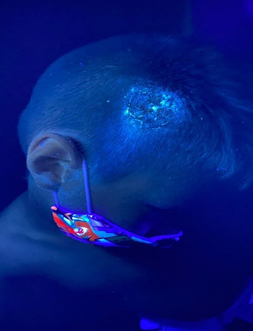
Tinea capitis in Children: A pandemic not yet eradicated.
Main Article Content
Abstract
Background: Tinea Capitis or Tinea Capitis is an almost exclusive childhood disease caused by one of the dermatophyte species, usually of the Microsporum and Trichophyton genera. Topic: among the risk factors associated with primary infection is using hairdressing equipment contaminated with microorganisms, contact with animals, or directly from person to person. The most relevant clinical data is the appearance of one or several alopecic plaques or pseudo alopecia with short and broken hairs, erythema, scaling, and occasionally the presence of papules and pustules. Methods: a narrative review. The identified articles came from the ScienceDirect, Scopus, PubMed, Medline, and Google Scholar databases, published between 1982 and 2021, with the following terms in the paper title: Tinea capitis, dermatomycosis in children, antifungals in children, children, diagnosis of Tinea capitis, epidemiology dermatomycosis. The words were related with connectors «AND», «OR». This review was carried out over six months, from August 2021 to January 2022. Results: the evolution of the infection can affect patients' quality of life, so microbiological confirmation is essential to allow proper treatment. Conclusion: management should be with oral medications for at least four weeks. Among the drugs used are griseofulvin, terbinafine, and itraconazole.
Downloads
Article Details

This work is licensed under a Creative Commons Attribution-NonCommercial-NoDerivatives 4.0 International License.
Creative Commons
License Attribution-NonCommercial-ShareAlike 4.0 International (CC BY-NC-SA 4.0)
You are free to:
Share - copy and redistribute the material in any medium or format.
Adapt - remix, transform, and build upon the material The licensor cannot revoke these freedoms as long as you follow the license terms.
• Attribution — You must give appropriate credit, provide a link to the license, and indicate if changes were made. You may do so in any reasonable manner, but not in any way that suggests the licensor endorses you or your use.
• NonCommercial — You may not use the material for commercial purposes.
• ShareAlike — If you remix, transform, or build upon the material, you must distribute your contributions under the same license as the original.
• No additional restrictions — You may not apply legal terms or technological measures that legally restrict others from doing anything the license permits.
References
Zuluaga A, Cáceres DH, Arango K, De Bedout C, Cano LE. Epidemiología de la tinea capitis: 19 años de experiencia en un laboratorio clínico especializado en Colombia. Infectio. 2016;20(4)225-230. DOI: https://doi.org/10.1016/j.infect.2015.11.004
Hay, R. Tinea Capitis: Current Status. Mycopathologia. 2017;182.(1-2), 87–93. DOI: https://doi.org/10.1007/s11046-016-0058-8
Messina, F., Walker, L., Romero, M. de las M., Arechavala, A. I., Negroni, R., Depardo, R et al. Tinea capitis: clinical features and therapeutic alternatives. Rev Argent Microbiol. 2021;19:S0325-7541(21)00011-0.
Kechia FA, Kouoto EA, Nkoa T, Nweze EI, Fokoua DC, Fosso S, Somo MR. Epidemiology of tinea capitis among school-age children in Meiganga, Cameroon. J Mycol Med. 2014;24(2):129-34. DOI: https://doi.org/10.1016/j.mycmed.2013.12.002
Abdel-Rahman SM, Farrand N, Schuenemann E, et al. The prevalence of infections with Trichophyton tonsurans in schoolchildren: the CAPITIS study. Pediatrics 2010;125: 966-973. DOI: https://doi.org/10.1542/peds.2009-2522
Monod M, Fratti M, Mignon B, et al. Dermatophytes transmitted by pets and cattle. Rev Med Suisse 2014;10:749-753.
Mirmirani P, Tucker LY. Epidemiologic trends in pediatric tinea capitis: a population-based study from Kaiser Permanente Northern California. J Am Acad Dermatol 2013;69: 916–921. DOI: https://doi.org/10.1016/j.jaad.2013.08.031
Zaraá I, Hawilo A, Aounallah A, Trojjet S, El Euch D, Mokni M et al. Inflammatory Tinea capitis: a 12-year study and a review of the literature. Mycoses. 2013;56(2)110-6. DOI: https://doi.org/10.1111/j.1439-0507.2012.02219.x
Mao L, Zhang L, Li H, et al. Pathogenic fungus Microsporum canis activates the NLRP3 inflammasome. Infect Immun 2014;82:882–892. DOI: https://doi.org/10.1128/IAI.01097-13
Takwale A, Agarwal S, Holmes SC, et al. Tinea capitis in two elderly women: transmission at the hairdresser. Br J Dermatol 2001;144:898-900. DOI: https://doi.org/10.1046/j.1365-2133.2001.04154.x
Aly R. Ecology, epidemiology, and diagnosis of tinea capitis. Pediatr Inf Dis J 1999; 18:180-185. DOI: https://doi.org/10.1097/00006454-199902000-00025
Brendan K. Superficial fungal infections. Pediatr Rev. 2012;33:e22-37. DOI: https://doi.org/10.1542/pir.33.4.e22
Negroni R, Arechavala A. Micosis superficiales de la piel y sus faneras. En: Lecciones de clínica micológica. 2 da ed Editorial Ascune; 2019. p.15-33.
Möhrenschlager M, Bruckbauer H, Seidl HP, Ring J, Hofmann H. Prevalence of asymptomatic carriers and cases of tinea capitis in five- to six-year-old preschool children from Augsburg, Germany: results from the MIRIAM study. Pediatr Infect Dis J. 2005;24:749-50. DOI: https://doi.org/10.1097/01.inf.0000172909.73601.dc
Panasiti V, Devirgiliis V, Borroni RG et al. Epidemiology of dermatophytic infections in Rome, Italy: a retrospective study from 2002 to 2004. Med Mycol 2007;45:57–60. DOI: https://doi.org/10.1080/13693780601028683
Fuller LC. Changing face of tinea capitis in Europe. Curr Opin Infect Dis 2009;22: 115-8. DOI: https://doi.org/10.1097/QCO.0b013e3283293d9b
Veasey J, Miguel B, Mayor S, Zaitz C, Muramatu L and Serrano J. Epidemiological profile of tinea capitis in São Paulo City. Anais Brasileiros de Dermatologia. 2017;92(2), 283-284. DOI: https://doi.org/10.1590/abd1806-4841.20175463
Proudfoot LE, Higgins EM, Morris-Jones R. A retrospective study of the management of pediatric kerion in Trichophyton tonsurans infection. Pediatr Dermatol 2011; 28: 655-7.
Pomeranz AJ, Fairley JA. Management error leading to unnecessary hospitalization for kerion. Pediatrics 1994; 93: 968-88. DOI: https://doi.org/10.1542/peds.93.6.986
Fuller LC, Child FJ, Midgley G, Higgins EM. Diagnosis and management of scalp ringworm. BMJ 2003; 326: 539–41. DOI: https://doi.org/10.1136/bmj.326.7388.539
Zhang R, Ran Y, Dai Y et al. A case of kerion celsi caused by Microsporum gypseum in a boy following dermatoplasty for a scalp wound from a road accident. Med Mycol 2011; 49: 90–3. DOI: https://doi.org/10.3109/13693786.2010.503196
Nenoff P, Kruger C, Schulze I et al. [Tinea capitis and onychomycosis due to Trichophyton soudanense: Successful treatment with fluconazole-literature review]. Hautarzt 2018; 69:737–50. DOI: https://doi.org/10.1007/s00105-018-4155-0
Burke WA, Jones BE. A simple stain for rapid office diagnosis of fungus infection of the skin. Arch Dermatol 1984;120:1519. DOI: https://doi.org/10.1001/archderm.1984.01650470125029
Head ES, Henry JC, MacDonald EM. The cotton swab technique for the culture of dermatophyte infections: its efficacy and merit. J Am Acad Dermatol 1984;11:797. DOI: https://doi.org/10.1016/S0190-9622(84)80455-7
Hiruma J, Ogawa Y, Hiruma M. Trichophyton tonsurans infection in Japan: epidemiology, clinical features, diagnosis and infection control. J Dermatol 2015;42: 245–249. DOI: https://doi.org/10.1111/1346-8138.12678
Robert R, Pihet M. Conventional methods for the diagnosis of dermatophytosis. Mycopathologia 2008;166:295–306. DOI: https://doi.org/10.1007/s11046-008-9106-3
Verrier J, Krahenbuhl L, Bontems O et al. Dermatophyte identification in skin and hair samples using a simple and reliable nested polymerase chain reaction assay. Br J Dermatol 2013;168:295–301. DOI: https://doi.org/10.1111/bjd.12015
Sugita T, Shiraki Y, Hiruma M. Real-time PCR TaqMan assay for detecting Trichophyton tonsurans, a causative agent of tinea capitis, from hairbrushes. Med Mycol 2006;44:579–81. DOI: https://doi.org/10.1080/13693780600717153
Verrier J, Monod M. Diagnosis of dermatophytosis using molecular biology. Mycopathologia 2017;182:193–202. DOI: https://doi.org/10.1007/s11046-016-0038-z
Ghannoum MA, Wraith LA, Cai B et al. Susceptibility of dermatophyte isolates obtained from a large worldwide terbinafine tinea capitis clinical trial. Br J Dermatol 2008;159:711–3. DOI: https://doi.org/10.1111/j.1365-2133.2008.08648.x
Arenas R, Toussaint S, Isa-Isa R. Kerion and dermatophytic granuloma. Mycological and histopathological findings in 19 children with inflammatory tinea capitis of the scalp. Int J Dermatol. 2006;45(3):215-9. DOI: https://doi.org/10.1111/j.1365-4632.2004.02449.x
Vazquez-Lopez F, Palacios-Garcia L, Argenziano G. Dermoscopic corkscrew hairs dissolve after successful therapy of Trichophyton violaceum tinea capitis: a case report. Australas J Dermatol. 2012;53:118–9. DOI: https://doi.org/10.1111/j.1440-0960.2011.00850.x
Richarz NA, Barboza L, Monsonis M et al. Trichoscopy helps to predict the time point of clinical cure of tinea capitis. Australas J Dermatol 2018;59: e298–e299. DOI: https://doi.org/10.1111/ajd.12830
Waśkiel-Burnat A, Rakowska A, Sikora M, Ciechanowicz P, Olszewska M, Rudnicka L. Trichoscopy of Tinea Capitis: A Systematic Review. Dermatol Ther (Heidelb). 2020; 10(1):43-52. DOI: https://doi.org/10.1007/s13555-019-00350-1
Seebacher C, Abeck D, Brasch J et al. Tinea capitis: ringworm of the scalp. Mycoses. 2007;50.218–26. DOI: https://doi.org/10.1111/j.1439-0507.2006.01350.x
Fuller LC, Barton RC, Mohd Mustapa MF et al. British Association of Dermatologists’ guidelines for the management of tinea capitis 2014. Br J Dermatol 2014;171:454–63. DOI: https://doi.org/10.1111/bjd.13196
Allen h, Honig P, Leyden J and McGinley K. Selenium sulfide: adjunctive therapy for tinea capitis. Pediatrics, 69 (1982), pp. 81-83. DOI: https://doi.org/10.1542/peds.69.1.81
Gupta AK, Mays RR, Versteeg SG et al. Tinea capitis in children: a systematic review of management. J Eur Acad Dermatol Venereol 2018;32:2264-74.
Gupta AK, Adam P, Dlova N et al. Therapeutic options for the treatment of tinea capitis caused by Trichophyton species: griseofulvin versus the new oral antifungal agents, terbinafine, itraconazole, and fluconazole. Pediatr Dermatol 2001;18:433-8. DOI: https://doi.org/10.1046/j.1525-1470.2001.01978.x
Chen X, Jiang X, Yang M et al. Systemic antifungal therapy for tinea capitis in children: An abridged Cochrane Review. J Am Acad Dermatol 2017;76:368-74.
B.E. Elewski. Cutaneous mycoses in children. Br J Dermatol, 134 (1996), pp. 7-11. DOI: https://doi.org/10.1111/j.1365-2133.1996.tb15651.x
Gupta AK, Mays RR, Versteeg SG et al. Tinea capitis in children: a systematic review of management. J Eur Acad Dermatol Venereol 2018; 32:2264–74.
Chen X, Jiang X, Yang M et al. Systemic antifungal therapy for tinea capitis in children: An abridged Cochrane Review. J Am Acad Dermatol 2017;76: 368–74. DOI: https://doi.org/10.1016/j.jaad.2016.08.061
F. Baudrez-Rosselet, M. Monod, S. Jaccoud, E. Frenk. Efficacy of terbinafine treatment of tinea capitis in children varies according to the dermatophyte species. Br J Dermatol. 1996;135(1996)1011-1012. DOI: https://doi.org/10.1046/j.1365-2133.1996.d01-1117.x
Higgins EM, Fuller LC, Smith CH. Guidelines for the management of tinea capitis. British Association of Dermatologists. Br J Dermatol. 2000;143(1):53-8. DOI: https://doi.org/10.1046/j.1365-2133.2000.03530.x
Gupta AK, Mays RR, Versteeg SG et al. Tinea capitis in children: a systematic review of management. J Eur Acad Dermatol Venereol 2018;32:2264-74. DOI: https://doi.org/10.1111/jdv.15088
Gubbins PO, Heldenbrand S. Clinically relevant drug interactions of current antifungal agents. Mycoses 2010; 53: 95-113. DOI: https://doi.org/10.1111/j.1439-0507.2009.01820.x
Solomon BA, Collins R, Sharma R, Silverberg N, Jain AR, Sedgh J, Laude TA. Fluconazole for the treatment of tinea capitis in children. J Am Acad Dermatol. 1997;37(2 Pt 1):274-5. DOI: https://doi.org/10.1016/S0190-9622(97)80141-7
Silverman R. Using oral antifungals safely. Contemp Pediatr 2001;18:9-11. DOI: https://doi.org/10.1046/j.1525-1470.2001.1988b.x
Blumer JL. Pharmacologic basis for the treatment of tinea capitis. Pediatr Infect Dis J.1999;18:191-9. DOI: https://doi.org/10.1097/00006454-199902000-00027
Hussain I, Muzaffar F, Rashid T et al. A randomized, comparative trial of treatment of kerion celsi with griseofulvin plus oral prednisolone vs. griseofulvin alone. Med Mycol.1999;37:97-9. DOI: https://doi.org/10.1046/j.1365-280X.1999.00199.x
Ginsburg CM, Gan VN, Petruska M. Randomized controlled trial of intralesional corticosteroid and griseofulvin vs. griseofulvin alone for treatment of kerion. Pediatr Infect Dis J. 1987;6:1084-7. DOI: https://doi.org/10.1097/00006454-198712000-00003
Proudfoot LE, Higgins EM, Morris-Jones R. A retrospective study of the management of pediatric kerion in Trichophyton tonsurans infection. Pediatr Dermatol. 2011;28:655–7. DOI: https://doi.org/10.1111/j.1525-1470.2011.01645.x
Mayser P. Treatment of dermatoses: Significance and use of glucocorticoids in fixed combination with antifungals. Hautarzt 2016; 67:732-8. DOI: https://doi.org/10.1007/s00105-016-3851-x
Schaller M, Friedrich M, Papini M et al. Topical antifungalcorticosteroid combination therapy for the treatment of superficial mycoses: conclusions of an expert panel meeting. Mycoses 2016;59:365-73. DOI: https://doi.org/10.1111/myc.12481
Stolmeier DA, Stratman HB, McIntee TJ, Stratman EJ. Utility of laboratory test result monitoring in patients taking oral terbinafine or griseofulvin for dermatophyte infections. JAMA Dermatol 2018; 154: 1409–16. DOI: https://doi.org/10.1001/jamadermatol.2018.3578
Patel D, Castelo-Soccio LA, Rubin AI, Streicher JL. Laboratory monitoring during systemic terbinafine therapy for pediatric onychomycosis. JAMA Dermatol. 2017;153: 1326-7. DOI: https://doi.org/10.1001/jamadermatol.2017.4483
Kramer ON, Albrecht J. Clinical presentation of terbinafineinduced severe liver injury and the value of laboratory monitoring: a critically appraised topic. Br J Dermatol .2017; 177:1279-84. DOI: https://doi.org/10.1111/bjd.15854
Cestari T, Manzoni A. Patología infecciosa. En: Larralde M, Abad E, Luna P, Boggio P, Ferrari B, editores. Dermatología Pediátrica. 3a ed. Ciudad Autónoma de Buenos Aires: Journal; 2021. p.174-266.


